Article Text

Statistics from Altmetric.com
Case report
A patient in his 20s presented to our trauma bay after sustaining multiple gunshot wounds to the left thorax. Upon arrival, the patient was hypotensive with a blood pressure of 95/58 mm Hg, heart rate of 129 beats/minute, respiratory rate of 29 breaths/minute, and oxygen saturation of 94% on 10 L non-rebreather. The patient was intubated for airway protection due to mentation and concern for his respiratory status. Focal assessment with sonography in trauma revealed a small anterior pericardial effusion. Initial chest X-ray showed a widened mediastinum and left hemopneumothorax with a bullet overlying the left hilum. Secondary survey and adjuncts created a puzzling picture. On the left side, the patient had three gunshot wounds in the mid-supraclavicular, superior mid-scapular, and posterior midaxillary regions (figure 1). Additional injuries included a left scapula fracture, left second and third rib fractures, and fractures of the anterior T1 and T2 vertebral bodies. On the right side, the patient had a palpable thrill at the right neck and supraclavicular region and was found to have a mid-shaft clavicle fracture and a large subclavian artery pseudoaneurysm with concern for active extravasation (figure 2). No injuries were identified in the abdomen or pelvis. After resuscitation and two units of whole blood in the trauma bay, blood pressure improved to 162/85 mm Hg.

X-ray revealing a metallic fragment overlying the left hilum with depiction of the bullet wounds. There is one anterior wound in the left mid-supraclavicular region (red dot). Two left-sided posterior wounds are depicted with green dots with one in the superior mid-scapular region and another along the posterior midaxillary region at about the level of the third rib.

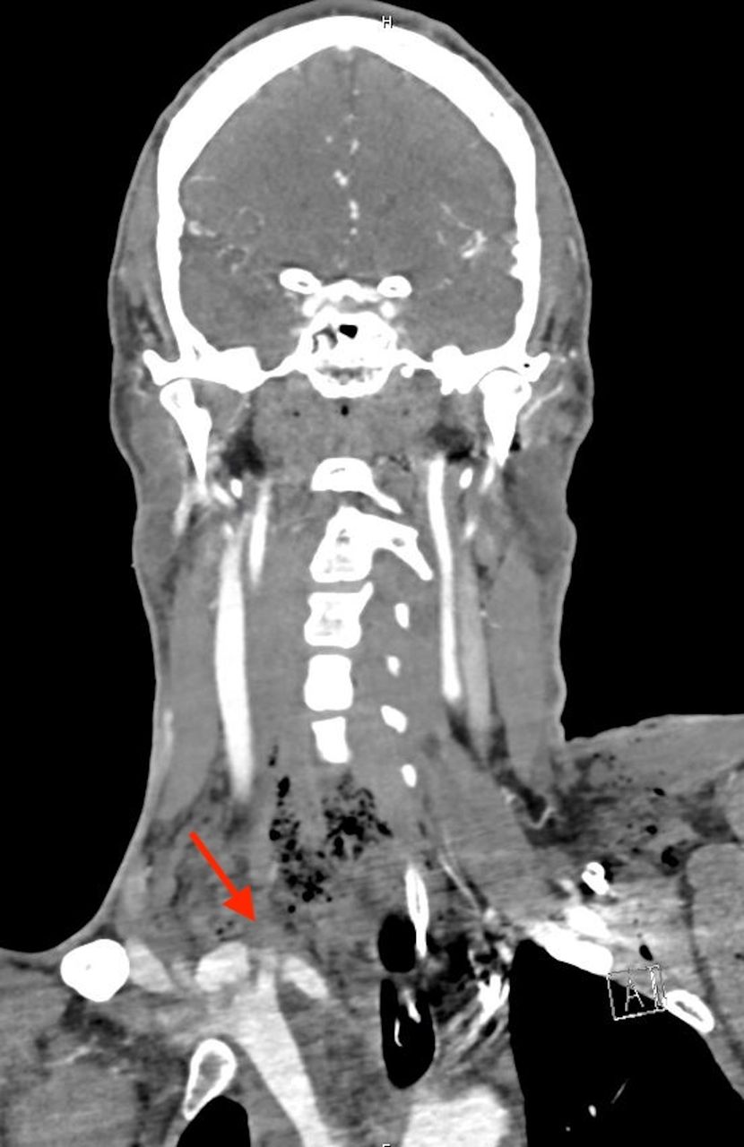
CT angiography coronal section showing a large pseudoaneurysm measuring 2.5×2.22 cm with probable extravasation.
What would you do?
Go to the operating room for mediastinal exploration.
Consult interventional radiology for endovascular management.
What we did and why
Correct answer: A
The clinical picture was consistent with all wounds and the retained bullet being located on the left side. However, the observed hematoma was on the medial aspect of right clavicle, prompting us to obtain further imaging to help delineate the injury pattern observed. Prior to transporting to CT scan for imaging, the patient denied any dysphagia. Furthermore, it was initially unclear that the gunshot wound was transmediastinal as we saw an injury to the proximal innominate artery on the right, but the missile appeared to be in the lung on the left and was not able to be clearly identified in the pulmonary artery due to missile artifact.
Despite the hemodynamic stability of the patient, he had hard signs of vascular injury with a palpable thrill at the neck. Due to suboptimal contrast filling, the exact location of vascular injury was not identifiable and upon discussion with radiology, they thought that there was injury to the proximal innominate artery that would be better accessed by median sternotomy.
In the operating room, there was no evidence of active hemorrhage and no identifiable injuries to the proximal great vessels so the patient was subsequently taken directly to interventional radiology where he was confirmed to have a right subclavian artery pseudoaneurysm with a complex arteriovenous fistula to the subclavian and innominate veins for which he underwent stent placement and multiple embolization procedures.
The bullet visualized on initial chest X-ray remained unmoved in a distal branch of the left pulmonary artery throughout the hospital course (figure 3). The patient denied any dyspnea, cough, or hemoptysis. Chest imaging was without evidence of ischemia or infarction of the lung parenchyma.

Three-dimensional reconstruction of CT angiography showing a metallic fragment lodged in the left pulmonary artery.
What would you do?
Open operative retrieval of the bullet
Conservative management and observation
Endovascular retrieval of the bullet
What we did and why
Correct answer: B
The decision was made to manage the bullet embolus conservatively as the patient was asymptomatic, without any evidence of right-to-left shunt, and there was no evidence of pulmonary infarction, abscess, or erosion in the bronchus. Furthermore, the risk of vascular injury or remigration more distally during manipulation may have outweighed any potential benefit of retrieval in this asymptomatic patient.
Discussion
Projectiles entering the body rarely follow a straight trajectory and instead take circuitous routes influenced by the patient’s position, gravity, hemodynamics, and the ability of the bullet to ricochet off internal structures.1 Consequently, the clinical presentation rarely correlates with what would be expected from the anticipated tract of the projectile. In our patient, a gunshot wound to the left thorax resulted in an injury to the right proximal great vessels, resulting in a traumatic arteriovenous fistula with subsequent embolization of the bullet to the right heart before reaching its destination in the left pulmonary artery.
These patients also present a therapeutic dilemma as there is no high-grade evidence to guide decision-making. Signs and symptoms that may warrant retrieval are those of pulmonary infarction, abscess, or erosion in the bronchus.1 2 Conservative management may be appropriate for patients who are both asymptomatic and without evidence of right-to-left shunting, as was the case for our patient.3–6
These patients may be at risk of long-term complications, but long-term outcomes are not routinely reported, and optimal outpatient follow-up has not yet been explored. Our patient was evaluated in the emergency department 2 months after injury for unrelated concerns. Chest X-ray revealed a stable bullet embolus with no evidence of complication. No further long-term follow-up is planned for the asymptomatic monitoring of the bullet embolus.
Ethics statements
Patient consent for publication
Ethics approval
This study involves human participants. This is a case report that is sufficiently anonymized and does not require IRB approval. Participants gave informed consent to participate in the study before taking part.
Footnotes
Contributors JLG was responsible for conception. NR was responsible for drafting of the article. JLG and HA were responsible for critical revision. All authors approved of the final article.
Funding The authors have not declared a specific grant for this research from any funding agency in the public, commercial or not-for-profit sectors.
Competing interests None declared.
Provenance and peer review Not commissioned; externally peer reviewed.