Article Text

Statistics from Altmetric.com
History
After a 48-year-old man noted the gradual onset of painless swelling of the entire left lower extremity over a period of several weeks, he went to the surgery outpatient clinic of a local trauma center. He stated that the significant swelling of the left lower extremity made it feel “heavy” and that he was having difficulty walking. He denied any recent prolonged history of immobility or trauma to the left lower extremity. In addition, he denied any history of deep venous thrombosis or surgery on the trunk or left lower extremity. On further questioning, he remembered that he had been a victim of a gunshot wound to the left groin in Mexico 26 years earlier. He was certain that he did not have surgery after this injury.
Examination
The patient was awake and alert with normal vital signs. A small circular scar, presumably from the old bullet wound, was noted in the left mid-groin 4 cm inferior to the inguinal ligament. The entire left lower extremity was edematous and slightly darker in color than the right lower extremity. There were normal pulses in the left common femoral artery and popliteal artery, whereas the left dorsalis pedis and posterior tibial pulses were present but difficult to palpate because of edema. There was no palpable thrill nor audible bruit in the left groin. Sensory and motor functions of the left lower extremity were normal.
Question
Based on the history and physical examination, the most likely diagnosis is:
Pseudoaneurysm of the left common femoral vein.
Thrombosis of the left femoral vein.
Congenital lymphedema (Milroy’s disease).
Traumatic femoral arteriovenous fistula.
Diagnosis
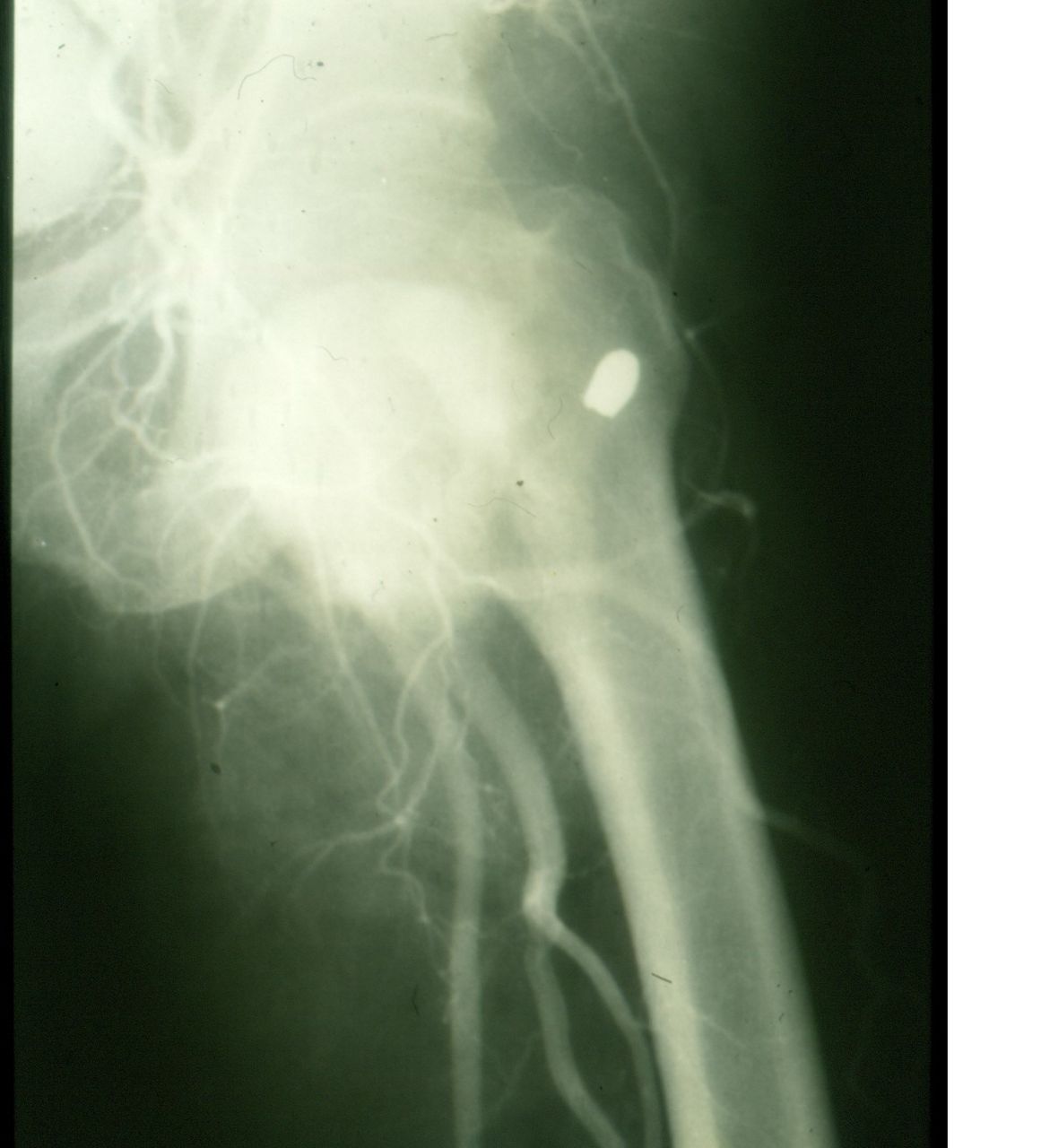
The patient was thought to have thrombosis of the left femoral vein, possibly related to the old gunshot wound to the groin. A left femoral arteriogram (patient cared for before availability of CT arteriography) documented that the patient had a large medial pseudoaneurysm of the left common femoral artery with intact flow into the superficial femoral and profunda femoris arteries (figure 1). No venous imaging was performed as there was presumed chronic thrombosis of the left common femoral vein secondary to compression from the pseudoaneurysm.
Arteriogram demonstrates a 26-year-old traumatic false aneurysm of the left common femoral artery.
Management
Although an endovascular stent graft would be considered to control the origin of the pseudoaneurysm in the modern era, the decision at the time the patient was cared for was to perform an open operation. The plan was that both arterial repair and possibly venous thrombectomy and reconstruction could be performed. The patient was typed and cross-matched for two units of packed red blood cells and received 1 g of a cephalosporin antibiotic intravenously in the preoperative area. The skin was prepared and draped from the umbilicus to bilateral toenails, and the left foot was placed in a sterile transparent bag. A longitudinal incision starting 2 cm superior to the inferior edge of the left inguinal ligament and extending 10 cm inferiorly was made over the left femoral arterial pulsations. Large venous collaterals encountered in the subcutaneous tissue were divided and ligated as needed for exposure. There was dense scarring in the area of the left common femoral artery as well as a medial pulsatile mass. A longitudinal incision 2 cm in length was made in the inferior edge of the left inguinal ligament to allow for exposure of the most distal left external iliac artery. There was less inflammation at this location, and a vessel loop was passed around the artery. The external iliac vein was thickened and collapsed at this location.
Distal arterial control with a vessel loop was obtained just proximal to the bifurcation of the left common femoral artery. The left common femoral vein was flattened as noted superiorly, but was encircled with a vessel loop as well. The area over the left common femoral artery and medial pseudoaneurysm was covered with dense scar tissue and large venous collaterals. After tedious and bloody dissection, 5000 units of unfractionated heparin was administered intravenously. Vascular clamps were then applied to the distal external iliac artery and to the distal common femoral artery. The medial 50% of the circumference of the mid-left common femoral artery was missing over 2 cm. A giant adjacent pseudoaneurysm cavity was noted to extend two-thirds of the way down the middle of the left thigh. After 15 min of irrigation and manual evacuation of old clot, the pseudoaneurysm cavity was relatively clean. A 4 cm segment of the left common femoral artery including the area of injury was excised, and an interposition graft was inserted. A completion arteriogram using fluoroscopy confirmed flow into the below-knee arteries of the left leg.
One option at this point was to expose the femoral vein where it was patent in the mid-left thigh. Then, a bypass graft of ringed polytetrafluoroethylene (PTFE) to the left external iliac vein could be performed through a left retroperitoneal exposure. Another option would be to see if the evacuation of the large pseudoaneurysm cavity would allow for further decompression of the left lower extremity as more venous collaterals developed. In light of the previous difficult dissection, the latter option was chosen and the incision was closed in layers.
Discussion
In contrast to patients who develop traumatic true aneurysms (tear of intima or intima-media with fusiform bulging of the artery; no blood outside of lumen) after blunt trauma, patients with penetrating wounds most often develop traumatic false aneurysms. These are full-thickness lacerations or loss of tissue in which blood is outside the lumen of the vessel. The original blood collection is labeled as an acute pulsatile hematoma. When operative or endovascular repair is delayed, the collection of blood may enlarge, there may be partial liquefaction of the surrounding clot, and there will be irritation of the surrounding tissues creating a pseudocapsule.1 The traumatic false aneurysm that results may cause any of the following: (1) pulsatile mass; (2) compression of the adjacent nerves or veins; (3) rupture with hemorrhage; or (4) distal arterial embolization.
The early history of vascular trauma surgery at the beginning of the 20th century is primarily about the delayed repairs of traumatic false aneurysms in military casualties.2 In the modern era, missed arterial injuries in the neck, trunk, and extremities occur in only 1%–4% of patients.3
The time from arterial injury until diagnosis of the traumatic false aneurysm varies, but was 52 years in one case report; however, the majority of patients are diagnosed early.4 In a review of the Vietnam Vascular Registry (includes false aneurysms and arteriovenous fistulas) by Rich et al 5 in 1975, nearly 53% of patients were diagnosed within 30 days of injury. The median delay until diagnosis in a civilian review of 28 patients by Feliciano et al 6 in 1987 was 10 days.
Spontaneous thrombosis of traumatic false aneurysms was noted to occur in 6.6% of wounded soldiers from World War II cared for by the late Harris B Shumacker, Jr, MD.7 For the remainder of patients, therapy depends on whether a critical artery is injured or not. Options for a non-critically injured artery are embolization, ligation of the artery around the wall defect (first described by Antyllus in Greece 21 centuries ago), and a modification of the obliterative aneurysmorrhaphy technique first described by Rudolph Matas in 1903.8
For traumatic false aneurysms in critical arteries, segmental arterial resection with an end-to-end anastomosis or insertion of an interposition graft is preferred. Depending on the artery involved and local expertise, the insertion of an endovascular stent graft is an attractive alternative.
Footnotes
Funding The authors have not declared a specific grant for this research from any funding agency in the public, commercial or not-for-profit sectors.
Competing interests None declared.
Patient consent Not required.
Provenance and peer review Commissioned; internally peer reviewed.