Article Text

Statistics from Altmetric.com
Case description
Our patient initially presented to the Urology service for robotic-assisted right simple nephrectomy due to complications from a ureteral stricture and recurrent obstructive pyelonephritis. In the operating room, the patient was placed in right-side-up flank position and access was obtained using the Veress needle at approximately the midclavicular line at the level of the costal margin. The initial pass of the Veress was questionable for bilious aspirate and the Veress removed. One more Veress attempt was made prior to plans to convert to Hasson, this time slightly medial. At the time of insufflation, the patient became hemodynamically unstable and was unable to be ventilated. Transthoracic echocardiogram confirmed a CO2 embolism. The incidence of CO2 embolism during laparoscopy is estimated to be 1 out of every 60 000 cases.1 Management included aborting the planned procedure, placing the patient in Trendelenburg/left lateral decubitus position to sequester air at the apex of the right ventricle, and providing supportive care through oxygenation and ventilation. The Veress was removed and the patient was positioned and stabilized by the anesthesiologist. The patient transferred to the ICU where they were resuscitated.
On postoperative day 3, the patient developed peritonitis and had an elevated white blood cell count of 16.3. CT scan of the abdomen revealed moderate volume intraperitoneal free fluid, with some fluid tracking around the gallbladder suggestive of bowel or biliary injury. Given the location of attempted Veress access, a gallbladder injury was suspected.
What would you do?
Broad-spectrum antibiotics and bowel rest.
Percutaneous cholecystostomy tube.
Return to the operating room for diagnostic laparoscopy and cholecystectomy.
What we did and why
Return to the operating room for diagnostic laparoscopy and cholecystectomy
Postoperatively, the patient developed a clinical examination suggestive of bile peritonitis. General Surgery returned to the operating room for diagnostic laparoscopy. On Hasson entry into the abdomen at the level of the umbilicus, we discovered a moderate volume of free-flowing bile. There were omental adhesions which were tethered to the gallbladder and the liver edge, which were taken down. These may have been related to prior surgical history of Roux-en-Y gastric bypass, or an inflammatory urologic process, and increased the risk for iatrogenic injury on Veress entry. The gallbladder was inflamed and ischemic (figure 1). We identified a small puncture injury near the fundus of the gallbladder, consistent with a hole made from Veress needle placement (figure 2). At this point, we were able to safely proceed with laparoscopic cholecystectomy. The patient recovered and remains with a nephrostomy tube to prevent further episodes of pyelonephritis, with plans to return for elective nephrectomy once fully recovered.
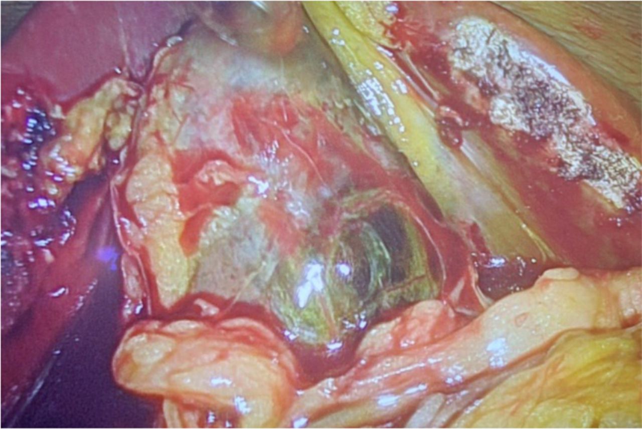
Inflamed and ischemic gallbladder secondary to trauma.

Iatrogenic perforation of gallbladder by a Veress needle.
Abdominal access in patients with prior abdominal operation or obese BMI may be technically challenging. Port placement for robotic right nephrectomy is performed with positioning at 90 degrees in left lateral decubitus (left side down). In obese patients, abdominal adipose tissue will migrate medially giving the appearance of an anterior lean when viewed straight on. This can distort the usual landmarks when estimating the midclavicular line. Access medial to the port line avoids entry into the retroperitoneum which can occur if the placement is too lateral. The proximity of the kidneys to other abdominal organs imparts risk of injury to these organs during operation. Vascular, intra-abdominal organ and urinary injuries have all been described. The right kidney is in immediate proximity to the liver, gallbladder, ascending colon and duodenum. Although rare, biliary complications including iatrogenic injury to the gallbladder have been reported and often necessitate cholecystectomy.2 Ischemic infarction of the gallbladder has been described in the setting of extramural arterial insufficiency, thrombosis or trauma.
There is no specific set of findings which are of diagnostic of gallbladder injury. Ultrasound may reveal heterogeneous hyperechoic blood adjacent to the gallbladder, as well as pericholecystic fluid. CT is more accurate in the setting of trauma for identifying blood or discontinuity of the gallbladder wall, as well as better at characterizing any concomitant adjacent organ injury.3 In the setting of bile peritonitis, preoperative imaging should not delay return to the OR for evaluation due to risk of sepsis and shock.
The management of gallbladder trauma (whether due to blunt, penetrating or iatrogenic injury) has traditionally been based on clinical symptomatology. Mild contusions or small lacerations may occasionally be managed conservatively. In the setting of larger lacerations, perforation or avulsion, surgical repair is warranted. Cholecystectomy is often the treatment of choice, with more conservative procedures such as percutaneous cholecystostomy reserved for patients who are poor surgical candidates.4 We advocate for immediate cholecystectomy once the injury is recognized (whether at the time of initial operation or on recognition postoperatively).
Footnotes
Contributors JDJ and AJS are the surgical residents involved in this case. JDJ is the first author and was primarily responsible for drafting the manuscript. AJS provided critical revisions. LMK is an Assistant Professor of Surgery in the Department of Surgery, Division of General Surgery; she was responsible for conceptualization and supervision of the manuscript, as well as critical revisions.
Funding The authors have not declared a specific grant for this research from any funding agency in the public, commercial or not-for-profit sectors.
Competing interests None declared.
Patient consent for publication Not required.
Provenance and peer review Not commissioned; externally peer reviewed.