Article Text

Statistics from Altmetric.com
History
A 27-year-old man presented to the trauma center with a gunshot would to the left groin exiting in the mid-right buttock. The emergency medical services technician accompanying the patient stated that blood loss was ‘1500–2000 mL at the scene’.
Examination
The patient was awake and alert with a heart rate of 124 beats/min, a blood pressure of 92/48, and a respiratory rate of 22 breaths/min. There were no peritoneal signs on abdominal examination, while there was normal tone and no blood on a rectal examination. He had a hematoma in the left groin which was oozing arterial blood, but no palpable pulses in the left lower extremity, and diminished motor and sensory function, as well. There was an exit wound in the mid-right buttock without bleeding or a hematoma.
Management
Large-bore intravenous access was obtained, an infusion of lactated Ringer’s solution was initiated, a pressure dressing was applied to the left groin, and a cephalosporin antibiotic was administered. The patient was taken to the operating room within 10 minutes of arrival in the trauma center.
Because of the proximity of the wound in the left groin to the inguinal ligament, a midline laparotomy was performed and vascular control of the left external iliac artery and vein was obtained. The left groin was then explored through a separate longitudinal incision, and vascular control of the left common femoral artery and vein was obtained, as well. The left external iliac artery under the inguinal ligament was noted to be transected, while the left external iliac vein in the same location was nearly transected.
Question
Based on the vascular injuries described, other presumed visceral injuries in the pelvis, and an admission base deficit of −15, your choice for management of the vascular injuries would be to:
Insert intraluminal shunts in both the artery and vein.
Ligate the vein, insert intraluminal shunt in the artery.
Ligate the vein, repair the artery using a graft from the internal iliac artery.
Ligate the vein, repair the artery using a plastic graft.
Management
Because of the patient’s hypotension, multiple intra-abdominal vascular injuries and the obvious presence of other abdominal injuries (to be described), the left external iliac vein and the left common femoral vein were both ligated. As the patient was now more hemodynamically stable, however, the transected ends of the left external iliac artery were debrided and an interposition graft of 8 mm externally supported polytetrafluoroethylene (PTFE) was placed between the left external iliac artery in the abdomen and the left common femoral artery in the groin.
An anterior wound in the bladder was seen along with extensive venous hemorrhage in the retropubic and left pelvic areas, presumably from ligation of the left external iliac and left common femoral veins. The surgical team then spent 30 minutes placing venous ligation stitches in these areas as well as applying fibrin glue. The anterior wound in the bladder was closed in two layers, while a posterior wound in the bladder was not repaired.
Laparotomy pad packing was placed in the retropubic and left pelvic areas, the open abdomen was covered with a silo, the incision in the left groin was partially closed (deep levels) to cover the PTFE graft, and the superficial layers of the left groin were packed open with dry gauze. At this point, the left leg was noted to be markedly swollen, though the left pedal pulses were still palpable. A left below-knee, two-incision, four-compartment fasciotomy was then rapidly performed by the attending surgeon and a second year fellow. The patient was transported to the intensive care unit with a systolic blood pressure of 142 mm Hg and a pulse of 160 beats/min after receiving 16 units of packed red cells, 1 unit of fresh frozen plasma, and 2 platelet packs (operation performed prior to massive transfusion protocols!).
Within 6 hours of surgery, the left thigh became swollen and tense. After a compartment pressure of 39–41 mm Hg was measured in the anterior compartment of the thigh, a fasciotomy of this compartment only was performed through an anterior incision in the intensive care unit.
The patient became hemodynamically stable during the next 48 hours, but with significant respiratory failure, and was returned to the operating room on postinjury day 3. Procedures performed included the following: (1) tracheostomy; (2) removal silo and intra-abdominal packing; (3) reopen anterior bladder repair and complete bladder neck repair and insertion of a Foley catheter by urology service; (4) resuspend bladder to pubic bone; (5) passage nasojejunal feeding tube; (6) closure of retroperitoneum over PTFE graft in left external iliac artery; (7) application silo to open abdomen; (8) closure of subcutaneous tissue and skin of left groin incision; and (9) fasciotomies of posterior and adductor compartments of left thigh.
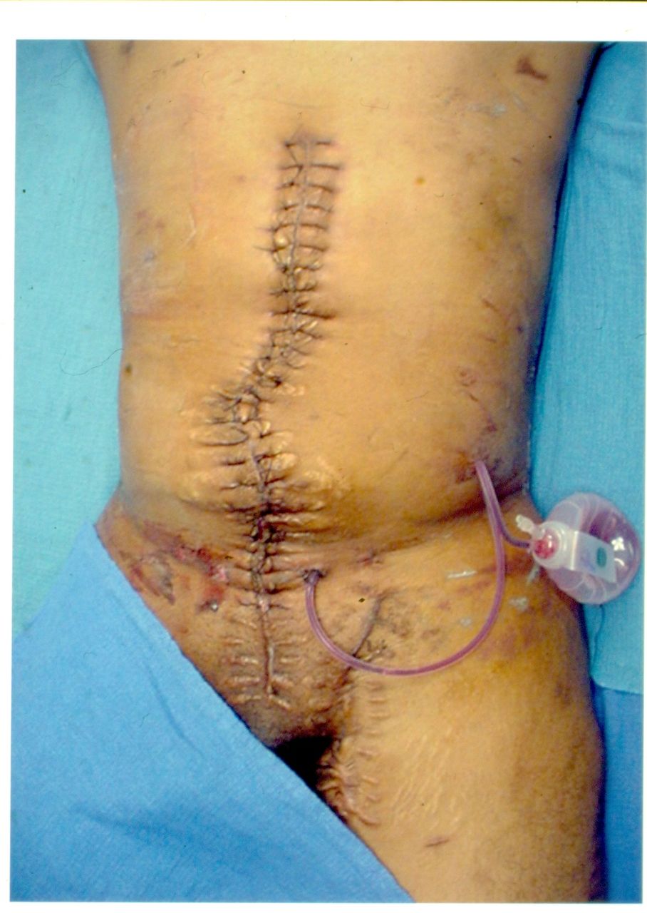
The patient subsequently had reoperations on postinjury days 4, 6, 10, and 30 to redo the repair of the bladder neck, reattach a torn silo to the abdominal wall, close the skin only of the abdominal incision, and close or cover the fasciotomy sites in the left leg and thigh. In addition, a filter was placed in the inferior vena cava on postinjury day 21. The patient was discharged from the trauma center 45 days after injury with all incisions healed (figure 1), 1–2+ edema of the left lower extremity (figure 2), and a protuberant abdomen secondary to the skin-only closure of the abdominal wall (figure 3). He was advised to use elastic wraps on his left lower extremity whenever sitting or standing until further notice.
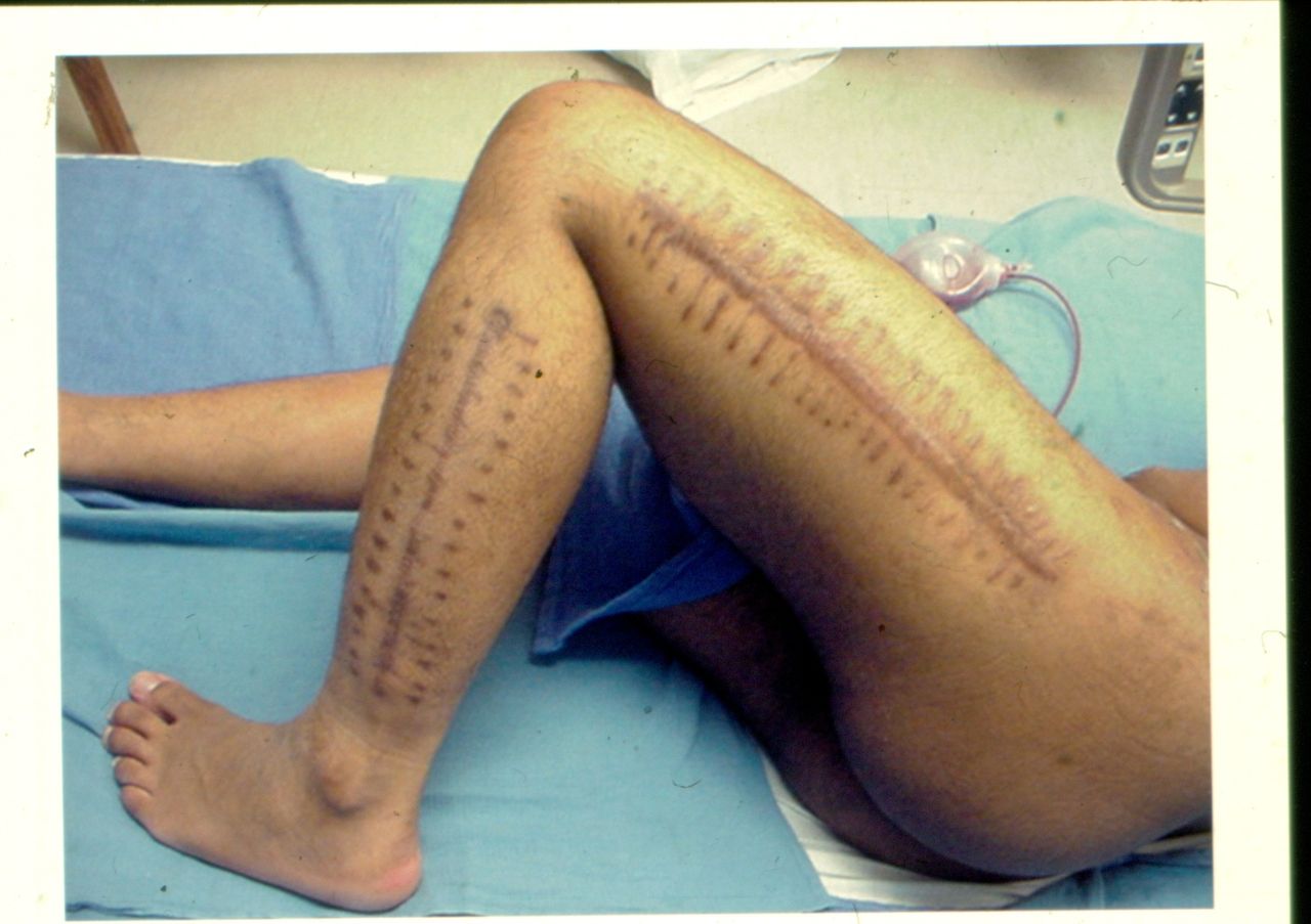
Well-healed lateral left thigh and calf fasciotomy incisions at time of discharge.
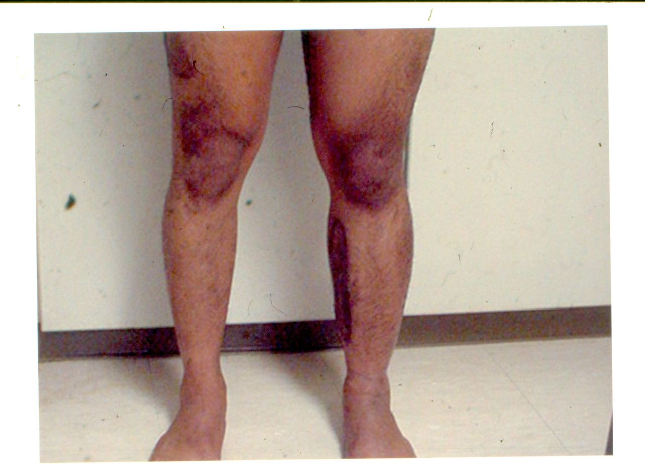
1–2+ edema of the left lower extremity at the time of discharge.

Protuberant abdomen at time of discharge after skin-only closure of abdominal wall during a series of ‘damage control’ operations.
The patient had an exploratory laparotomy, lysis of adhesions, and components separation closure of the abdominal midline incision 255 days after injury to correct the protuberant abdomen (figure 4). At that time he was noted to have persistent 1–2+ edema of the left lower extremity, but had ceased applying elastic wraps.
Flat appearance of abdomen after components separation closure of abdominal wall 255 days after injury.
Discussion
The original indications for ‘damage control’ approaches in abdominal trauma include hypothermia <35°C, a metabolic acidosis with a pH <7.2 or a base deficit <−15, and/or a coagulopathy with an international normalized ratio or partial thromboplastin time >50% of normal reflected intraoperative concerns about ‘physiologic exhaustion’.1–4 The number of indications has increased substantially since then, and the technique is now one of the most overused in the field of abdominal trauma.5 6 The patient described, however, with a history of significant preoperative blood loss, profound hypotension at admission, the presence of combined abdominal vascular injuries, and other intra-abdominal visceral injuries would fit everyone’s indication for ‘needs damage control’.
The operative approach to patients with vascular injuries posterior or adjacent to the inguinal ligament has been discussed in a previous ‘Case of the Month’ in Trauma Surgery and Acute Care Open.7 Using a midline abdominal incision to obtain intra-abdominal retroperitoneal control of the external iliac vessels and a separate longitudinal groin incision to obtain control of the common femoral vessels ‘saves’ the overlying inguinal ligament. The inguinal ligament will then be another layer of coverage over any vascular repair, graft, or ligation in the retroperitoneum and should help protect any of these from a postoperative infection in superficial soft tissue of the groin.
In the modern era, many surgeons would have inserted temporary intraluminal shunts into the external iliac artery or both artery and vein in light of the patient’s profound hypotension and combined major vascular injuries. It is important to recognize, however, that the larger shunts needed to fit the common or external iliac artery may not be available in every operating room (Pruitt F3 Outlying and Inlying Carotid Shunts with T-Ports, 10F, LeMaitre, Burlington, Massachusetts; Bard Burbank Carotid Bypass Shunt, 18 F tapered to 12 F, and Bard Javid Carotid Bypass Shunt, 17 Fr tapered to 10 F, Bard Peripheral Vascular, Tempe, Arizona). In such a circumstance, large-bore intravenous tubing or an appropriately sized thoracostomy tube may be used as a shunt.
Short-term and long-term data document that ligation of an injured external iliac vein is generally well tolerated in young and otherwise healthy trauma patients.8 Important principles of management after this ligation include the following: (1) elevation of the entire ipsilateral lower extremity when the patient is supine or sitting (unless compartment syndromes are impending); (2) elastic wraps whenever the patient is sitting or standing if any postoperative edema is present; and (3) a vigorous walking program once the patient has recovered from a laparotomy.
The disadvantages of ligation of the external iliac vein (and common iliac vein and inferior vena cava) were well demonstrated in the patient described. First, there was severe intraoperative bleeding from every pelvic venous collateral, including those around the injured bladder, that mandated extensive suture ligation. Second, the patient needed both a four-compartment fasciotomy of the leg during the first operation and a three-compartment fasciotomy of the thigh in the postoperative period. While the need for thigh fasciotomy is uncommon in injured patients, a compartment pressure exceeding capillary pressure (30–35 mm Hg) which is likely to be prolonged should always prompt consideration for performing the procedure. This is particularly true when diffuse swelling is due to ischemia/reperfusion or venous ligation rather than an isolated fracture. And, third, the patient developed early postoperative and chronic edema of the left lower extremity.
The choice of a substitute vascular conduit in wounds of the common iliac, external iliac, or common femoral artery at a first operation or after removal of a temporary intraluminal shunt at a reoperation depends on hemodynamic status, presence of associated gastrointestinal injuries, and, often, the size of the contralateral greater saphenous vein. Replacement of these vessels requires an 8–10 mm conduit, and the greater saphenous vein will usually not dilate to this size after retrieval. In the patient described, as in all critically injured patients in the author’s experience, creating a spiral vein or panel vein graft (a 45-minute endeavor) has no appeal. Without an associated gastrointestinal injury, a ringed PTFE graft of appropriate size was chosen for arterial replacement under the inguinal ligament. Mandatory precautions to avoid infections and maintain patency in synthetic conduits after vascular trauma include the following: (1) complete coverage of the prosthesis with well-vascularized tissue; (2) administration of perioperative antibiotics; (3) early administration of rectal or oral daily aspirin 81 mg/day; (4) cessation of smoking; (5) avoid sitting for long periods when the prosthesis crosses the hip or knee joint; and (6) a vigorous walking program after recovery from operation.
Ever since the admonition against the use of ‘prosthetic material’ in military vascular wounds in Vietnam by Norman Rich and the late Carl Hughes in 1972, there has been a reluctance during subsequent military conflicts to counter this advice.9 A novel approach to overcome this concern, however, has been reported from the experience in Iraq and Afghanistan.10 In patients with vascular trauma in an extremity and in whom there were ‘limited noninjured vein conduits’, PTFE grafts were inserted as a temporary solution to maintaining arterial flow. This allowed for ‘patient stabilization, transport to a higher echelon of care’, and later elective revascularization (PTFE graft removed) ‘with remaining limited autologous vein’.
Data availability statement
There are no data in this work.
Footnotes
Funding The authors have not declared a specific grant for this research from any funding agency in the public, commercial or not-for-profit sectors.
Competing interests None declared.
Provenance and peer review Commissioned; internally peer reviewed.