Article Text

Statistics from Altmetric.com
History
A 29-yearold man presented to the trauma center with a single gunshot wound to the right costal margin.
Examination
The patient was alert and responsive on arrival, but became increasingly lethargic throughout the assessment. His initial vital signs were a heart rate of 100 beats per minute, a systolic blood pressure of 80 by palpation and a respiratory rate of 20/min. His abdominal exam was significant for a single gunshot wound to the right costal margin at the midclavicular line, and his abdomen was distended and tense to palpation.
Question
The most appropriate first step in management of this patient in addition to resuscitation is:
Massive transfusion and abdominal CT scan.
Surgeon-performed FAST (focused assessment for the sonographic evaluation of trauma) examination.
Transfer to operating room.
Emergent hepatic arteriogram.
Management
A massive transfusion protocol was initiated, packed red blood cells were administered, and the patient was taken directly to the operating room where he underwent endotracheal intubation. After the administration of a cephalosporin antibiotic, a midline exploratory laparotomy was performed. A significant hemoperitoneum was evacuated. After evisceration of the small bowel to the right, the infrarenal abdominal aorta was cross-clamped because of profound hypotension. A large right retroperitoneal hematoma was exposed by a right-sided medial mobilization maneuver. After visualizing a 5 cm longitudinal anterior split in the infrarenal inferior vena cava (IVC) with a corresponding 2 cm posterior perforation just above the confluence of the common iliac veins, vascular cross-clamps were applied to both common iliac veins and the infrarenal IVC. There was still significant back-bleeding from the lumbar veins. After an attempt to repair the significant anterior laceration in the IVC, it became obvious that the IVC would be significantly narrowed. In addition, the patient remained profoundly hypotensive. At this point, both common iliac veins, the infrarenal IVC just above the perforations, and several lumbar veins were ligated. Other injuries found on rapid exploration of the abdomen included the following:
Grade I injury to the edge of the right lobe of the liver; several perforations in the duodenum including a transection at the junction of D2 and D3; and a Grade II injury to the neck of the pancreas. The duodenal perforations and transection were stapled closed, the periduodenal area and right retroperitoneum were packed, and temporary abdominal coverage was applied. Prior to leaving the operating room, compartment pressures in both legs were measured and were noted to be >35 mm Hg. Bilateral two-skin incision four-compartment fasciotomies were rapidly performed in both legs. At the end of the first operation, the patient had received 26 units of packed red blood cells, 8 units of fresh frozen plasma, 10 units of platelets, and 10 units of cryoprecipitate.
The patient was returned to the operating room the next day for pack removal, washout, oversewing of the third portion of the duodenum, repacking of the right retroperitoneum, and repeat temporary abdominal coverage. Later the same day, the patient's thighs were noted to be swollen and tense.
Question
Appropriate release of the compartments in the thigh can be performed through the following:
A single medial incision to release the medial (adductor) compartment.
Medial and lateral incisions to release the anterior (extensor), medial (adductor), and posterior (flexor) compartments.
Anterior and posterior incisions to release the extensor and flexor compartments.
S-shaped incision to release the anterior, lateral, and posterior compartments.
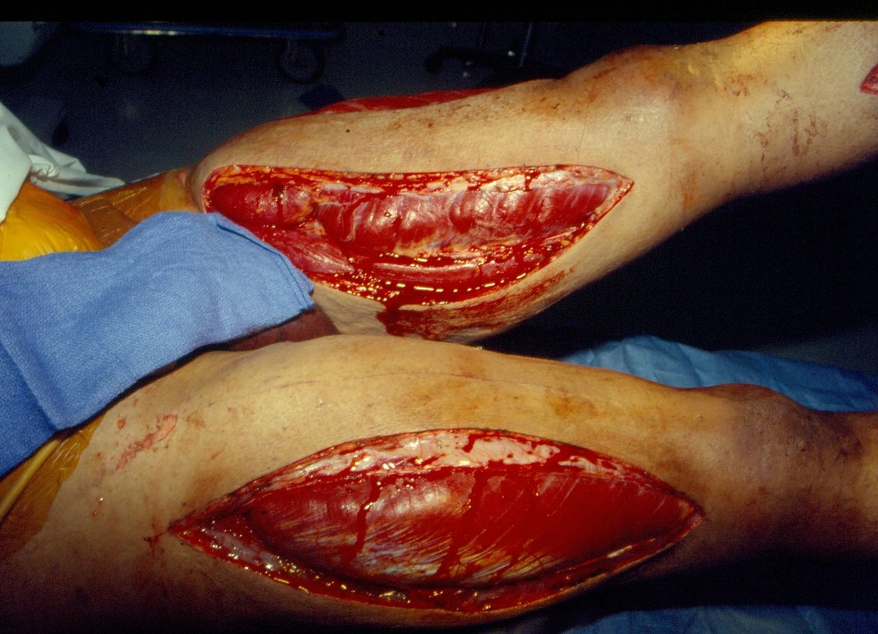
Due to the concern for compartment syndromes in both thighs, the patient was returned to the operating room and bilateral two-skin incision (medial and lateral) three-compartment thigh fasciotomies were performed1 (figure 1). On post-injury day 2, he was returned to the operating room for a fourth operation. A handsewn end-to-end duodeno-duodenostomy was performed between the second and third portions of the duodenum (figure 2). In addition, an omental pedicle was placed over the Grade II injury of the neck of the pancreas, a pyloric exclusion was performed with a #1 polypropylene suture, and a handsewn antecolic gastrojejunostomy restored gastrointestinal continuity. Finally, a jejunal feeding tube was placed (Witzel), and the abdomen was covered with an absorbable mesh. On post-injury day 18, split-thickness skin grafts (STSG) were placed on all leg fasciotomy and the left lateral thigh fasciotomy sites (figure 3). Non-healing STSGs on fasciotomy sites were excised and Alloderm grafts were placed on post-injury day 31. On post-injury day 43, a STSG was placed over the open abdomen, and the patient was discharged home without edema of the lower extremities on post-injury day 49 after seven operations (figure 4).

Bulging of thigh muscles after bilateral fasciotomy at third operation.

End-to-end duodeno-duodenostomy between second and third portions of duodenum at fourth operation.

Split-thickness skin graft applied to fasciotomy site on left lateral thigh at fifth operation.

Patient at discharge on post-injury day 49 after seven operations.
Question
All of the following are appropriate management options for significant infrarenal IVC injuries with exsanguination except:
Dacron or ringed *PTFE (polytetrafluoroethylene) interposition graft.
Cavo-atrial bypass graft.
Primary repair.
Ligation.
Correct answer is B. This option would only be used after ligation of an injury to the juxtarenal or suprarenal infrahepatic IVC.
Discussion
Mortality rates from IVC injuries have risen in recent years despite the availability of resuscitative endovascular balloon occlusion of the aorta, damage control operative techniques, and, on occasion, endovascular balloon occlusion or stent placement.2 In one study in 2014, the mortality rate was 47.8%.3 Crucial to the outcome is rapid identification and repair or ligation of the area of injury. Exposure of the entire infrahepatic IVC to the confluence of the common iliac veins involves mobilization of the right colon and duodenum medially (right-sided medial mobilization maneuver). If significant bleeding impedes visualization of the injury to the IVC, spongesticks with folded sponges can be used to compress the IVC above and below the area of injury. Small anterior or lateral perforations of the IVC can be controlled by the application of a Satinsky vascular clamp. Larger anterior or lateral perforations or blunt lacerations can be controlled by the application of sequential Judd-Allis clamps before suturing. Combined large anterior and posterior perforations or a significant longitudinal laceration may require the application of DeBakey angled vascular or long curved aortic clamps around the area of injury. Complete occlusion of the infrarenal IVC in the patient with hypovolemia, however, will increase hypotension due to decreased venous return. Therefore, the infrarenal abdominal aorta should always be cross-clamped simultaneously. Perforations at the renal vein–IVC junction (juxtarenal) or at the confluence of the iliac veins are particularly challenging.4 With the former, medial rotation of the kidney can allow for better control of the renal vein–IVC junction or expose a posterior perforation. Another option is to cross-clamp the infrarenal and suprarenal IVC as well as both renal veins around the area of the injury. With wounds to the common iliac vein–IVC junction, the internal iliac artery on the side of injury can be divided and ligated to allow lateral or medial retraction of the overlying common iliac artery. An alternate approach is temporary cross-clamping and division of the right common iliac artery followed by left lateral mobilization of the aortic bifurcation.5 This allows for complete exposure of the common iliac vein–IVC junction (as well as the posterior distal infrarenal abdominal aorta). After the venous (or aortic) repair, Fogarty balloon catheters are passed into the arterial tree through the distal end of the right common iliac artery and the artery is reconstituted with an end-to-end anastomosis. In a patient with physiologic exhaustion and a large IVC injury or when the IVC injury would require an interposition graft, the infrarenal IVC should be ligated. This damage control approach is associated with higher postoperative complication rates than primary repair or patch closure, as expected. A retrospective propensity-score matching analysis of all IVC injuries from 2010 to 2014 in the National Trauma Data Bank found no mortality benefit to IVC ligation versus repair and higher associated rates of pneumonia, deep venous thrombosis, pulmonary embolus, and extremity compartment syndromes in the ligation group.6 If ligation of the infrarenal IVC is performed, the patient should be resuscitated with blood and even crystalloid solutions and monitored closely for the development of a compartment syndrome (as in the patient described) and deep venous thrombosis in the lower extremities. Despite the higher short-term complication rate after ligation of the IVC, long-term sequelae are modest in young patients. This is especially true if the lower extremities are elevated while the patient is at bed rest and elastic wraps are applied whenever the patient ambulates during hospitalization and for 3 months as an outpatient. In a 2010 retrospective review of 22 patients who underwent ligation of the infrarenal IVC at Grady Memorial Hospital, nine (41%) survived to discharge.7 Within that group, none had more than trace edema of the lower extremities at the time of discharge from the hospital. On long-term follow-up of seven patients, none had any progression of edema.
Endovascular approaches have been described in animal studies and patient case reports of both temporary balloon occlusion and definitive stent-graft management of IVC injuries. A swine model using endovascular occlusion of the suprahepatic IVC for a juxtahepatic IVC injury demonstrated an 83% survival in the control arm at 1 hour after injury.8 A more recent report in a swine model described the use of a stent graft for temporary control of uncontained hemorrhage from the IVC and noted that there was control of hemorrhage in all seven animals.9 In patient case reports, stent grafts have been inserted into hemodynamically stable patients with contained retroperitoneal hematomas. To date, there have been no multicenter trials or large series demonstrating successful endovascular management of bleeding from an injured IVC.
Footnotes
Funding The authors have not declared a specific grant for this research from any funding agency in the public, commercial or not-for-profit sectors.
Competing interests None declared.
Patient consent for publication Not required.
Provenance and peer review Commissioned; internally peer reviewed.